Title Image
You are in:  / Home  /  OCULAR SURFACE UNIT  /  OCULAR SURFACE UNIT

OCULAR SURFACE UNIT

In response to the study and solution of axonal degeneration in the cornea, our research projects over the course of 2016 were:

PROJECT 1.    AXONAL DEGENERATION ARISING FROM ENDOGENOUS FACTORS: AGING AND NEURODEGENERATIVE DISEASES

A)    AGING

The natural aging process affects all tissues in a progressive manner. The nervous system is no exception. Older people lose their sight, hearing, smell and even taste. But they also find their sense-performance decreasing on the whole. Fine touch, temperature and pain are senses that undergo changes with aging. In many cases a usually innocuous stimulus becomes a painful and uncomfortable sensation, which we call allodynia. The common view is that as we age, we lose a considerable amount of sensory receptors and that this is the cause for sensory dysfunction. Some pathologies such as diabetes can accelerate the degeneration of nerves and aggravate the process, indeed even causing chronic and painful ulcers on the skin.

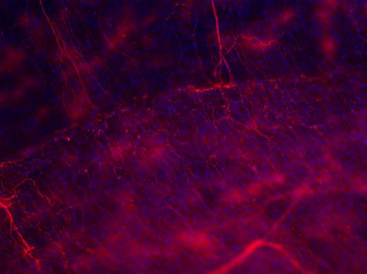
However, not all structures appear to be equally vulnerable to aging. In our laboratory we have studied sensorial innervation of the ocular surface in aging mice and have focused on the analysis of cold-sensitive receptors, due to their special importance in the regulation of tear-secretion. We have identified and characterised both functionally and molecularly, cold-receptor neuron populations that feature the TRPM8 ionic canal in the cornea. Furthermore, these two sorts of neurons have different functions. The first type plays a regulatory role in the production of tear secretions and we believe that it contributes to the blocking of pain by inhibiting other poly-modal neurons. The second kind displays typical characteristics of a classical nociceptor. With focus being solely on these two nerve-fibre types of the cornea, we observed that aging affects each of them in a different manner, with interesting singular degenerative processes in the cornealafferents: degeneration linked to aging in the cold fibres takes place almost exclusively in the first of the two fibre types, while the other type is not significantly affected. The selective disappearance of the “regulator” kind of TRPM8 fibres, is linked to dysfunction of the tear-duct. The “nociceptor” type of the TRPM8 fibres remains present in older animals display pain and inflammation markers. In accordance with our hypothesis, the latter would be responsible for the appearance of bothersome and painful sensations that go in hand with tear-duct dysfunction, in patients of advanced age. Similarly, in aging animals we find an electrical activity in a population of cold-terminal population that appears similar to that of the regulator fibres (which in normal conditions has a constant basal activity level) but with a response activity that is higher and more prolonged, similar to that of poly-modal fibres.

In this way, we have been able to directly link selective loss of nerve fibres in the cornea to tear-duct dysfunction associated with aging, in a model that accurately reflects the pathological signs of the dry eye: tear-duct dysfunction, inflammation and pain.
The dual activity of the TRPM8 canal defines it as a therapeutic target for solving neuropathic and inflammation-related pain.
On the other hand, understanding the reason for which a type of neuron so restricted displays greater vulnerability to degeneration, opens avenues for the design of neuro-protective treatments.

B) NEURODEGENERATIVE DISEASES

In tight correlation with the aging process, we find neurodegenerative diseases. At the present time it is believed that neurodegenerative diseases are systemic illnesses with symptomatology and involvement predominantly in the central nervous system. Clinical evidence defining a neuro-degenerative illness usually includes cognitive or severe motor deficiency, detected at an advanced age. However, the start and progress rate of the illness occur at a much earlier stage in life. Neural and synaptic plasticity processes of the brain obscure the progress of the pathology and maintain cognitive and motor functionality until a later point of inflexion. In some neurological diseases such as Parkinson’s, high levels of α-sinnucleine (a bio-marker for the illness) present in colon biopsies, up to 15 years before the appearance of the earliest motor symptoms.

In Alzheimer’s disease, peripheral neuropathies can be detected in skin biopsies from patients. In animal models of neurodegenerative diseases we have observed that the eye is extremely vulnerable to systemic illnesses, such as for example in the case of hepatic encephalopathy. Both the retina and the innervation of the cornea may show early-onset signs of nervous degeneration, previous to the appearance of cognitive deficiencies typical in neurological disease. In our case, we have observed that mice with neurodegenerative diseases exhibit certain neuropathies in the cornea. The dystrophic axons, in this case, show similarities both in “dying back” processes and in anterograde deteriorations similar to Wallerian degeneration.

This blend of mechanisms may point to a direct neuronal affectation, which is in turn sustained by the presence of the hyper-phosphorylated TAU protein in the soma and the axon.

content_figura1_1

Figure 1: View of the dystrophic nervous fibres in the cornea of a mouse APP/PS1. Note the elevated quantity of varicosities.

PROJECT 2.    REGENERATION IN INDUCED NERVE LESIONS: MECHANISMS FOR NERVE REGENERATION AND NEURO-PROTECTION.

A)    STUDY OF NERVE REGENERATION IN EXPERIMENTAL SURGICAL LESIONS.

Employing the proprietary model for cornea lesion by laser excision (photorefractive surgery or PRK) we can produce a lesion by axotomy of the entire sub-basal nerve plexus of the cornea. Over the course of 3 months the morphological parameters of regeneration are studied, their relation to scarring of the tissue and the recovery of their function previous to the lesion. These results will be very valuable in devising treatments directed towards corneal repair and the relief of post-surgical pain. The implication in nervous regeneration of other cell types of the non-nervous variety, such as the glia, fibroblasts and inflammatory cells, is mostly unknown both in literature and in clinical practice. It is also important to know the change in the extracellular environment during regeneration and its involvement in cases of aberrant regeneration. The setting of basal parameters in regeneration serves as the origin for experimentation in different regenerative approaches.

The rate of regeneration in the cornea depends on the presence of an appropriate substrate for the emission of neurites and the presence of terminal targets in the receptors. In our experiments, we generally found no axons in regeneration previously to the complete re-epithelialisation of the damaged area of the tissue, which usually takes place between 3 and 4 days after the lesion. After three months of recovery, the innervation of the corneal surface has a normal appearance, even featuring an apical nervous vortex. The electro-physiological activity in this stage is normal.

content_figura2_2

Figure 2: View of the peripheral region of the corneal lesion in which the branching and elongation of the axons in regeneration towards the cornea centre (located in the lower right corner of the image), is observed.

B)    STUDY OF THE NERVE LESION INDUCED BY LIGHT RADIATION.

Besides the traumatic or surgical nerve lesions massively affecting the sensory innervation of the cornea, other many external factors may be considered as being exogenous degenerative factors in the cornea. Light radiation of a certain wave length, for example, has been described as inducing or enhancing of degeneration and death in retinal cells, especially if the retina is in a pathological state.

In our lab we have shown the cornea to be also affected by light radiation and that it preferentially damages nervous constituents. Specifically, we have recognised degenerative structures in the axons coinciding with degenerative “dying back” mechanisms. Our hypothesis, which is shared by Dr. Neville Osborne, is that the light interferes with normal mitochondrial respiration chain activity, reducing its capacity in the face of oxidative stress.

The accumulation of reactive species in the oxygen (ROS) causes deterioration in the mitochondrial function necessary for normal neuron function. In the axon’s local environment, microtubules become unstable as a result of mitochondrial deficit and the phosphorylation of TAU, becoming grouped in the thickening of the membrane (varicosities) throughout the axon. Vesicular traffic along the axon becomes interrupted and finally the plasma membrane of the axon is reabsorbed, giving way to degenerative spherules. The function of the nerve fibre is lost.

Once we understand this scenario, the following step will be the application of neuro-protective treatments for inhibiting or preventing exogenously induced lesions. An effective neuron protection would allow for the rescue of fibres being affected by the aging process, for example.